

Western Blot
Western blot is a well-established and widely used technique for the detection and analysis of proteins. The method is based on the construction of an antibody-protein complex with specific antibodies that bind to proteins immobilized on a membrane. In the last decade, The Fusion has permit great improvements in detection methods and software that have made quantitative Western blot safer and more sensitive and efficient.
A variety of detection systems, based on chemiluminescence, fluorescence, or radioisotopic detection are available. Radioisotopic reagents have seen a steady decline due to safety issues with handling radioactive isotopes. Consequently, enzyme-based chemiluminescence and direct fluorescence have now become the systems of choice, offering both high sensitivity and wide dynamic ranges.
Western Blotting Detection Methods
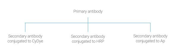
Chemiluminescence detection in Western blotting is based on antibodies conjugated to the enzyme horseradish peroxidase (HRP) that catalyzes the oxidation of luminol in the presence of peroxide, and results in light emission.
Normally the reaction produces low intensity emission of light at 428 nm. However, in the presence of enhancers, such as, modified phenols and, especially p-iodophenol, the emitted light is enhanced up to 1000-fold. This simplifies detection and increases the sensitivity of the reaction; the whole process is known as enhanced chemiluminescence (ECL). The intensity of signal is a result of the number of reacting enzyme molecules and is thus proportional to the amount of antibody, which is related in turn to the amount of protein on the blot.
Western blot imaging process
- Allow the ECL to equilibrate to room temperature before opening the vials
- Mix an equal volume of ECL solutions A and B, allowing sufficient total volume to cover the membranes. A volume of 0.1 ml/cm2 of membrane is required
- Drain the excess washing solution from the washed membranes and place them, protein side up on the Fusion blot sample tray. Pipette the mixed detection reagent onto the membrane
- Incubate for 1 min or more according to the ECL protocol. The incubation time could reach 5 minutes. at room temperature
- Place the sample tray in the Fusion system and operate according to instructions. Choose automatic exposure time and capture the image
Choose exposure times according to expected signal intensity if you are familiar with your system. Recommended is to begin with automatic exposure time and then adjust the time to find the optimal exposure. Alternatively, an increment function can be used in which the camera captures images at predetermined time points during a given time.
Auto-exposure in the ECL time laps environment
As the light intensity change with time, the auto-exposure is the perfect imaging mode for chemiluminescence. It compensate the light change by increasing or decreasing the calculated exposure time and optimize the image dynamic.

